The NPN Lab seeks to understand neural circuits and their dynamics in disease states and to develop precise neuromodulation strategies to treat neurological and neuropsychiatric disorders. We combine direct recordings from the human brain, mechanistic studies in animal models, and computational modeling to investigate disease-relevant neural activity and develop individualized approaches to modulate dysfunctional brain circuits.
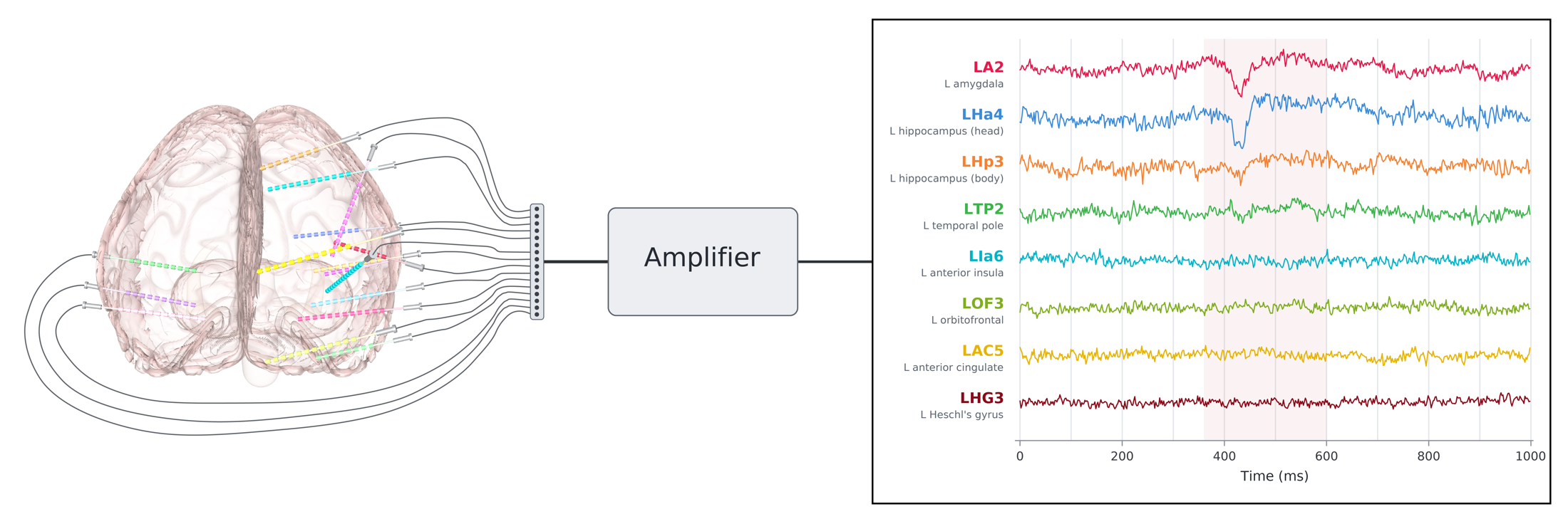
Stereo-electroencephalography (sEEG) is a technique in which electrodes are temporarily implanted in multiple brain regions for 7–14 days to monitor seizures, allowing electrical activity to be recorded directly from the human brain with high spatial and temporal resolution over many days. Through our neurosurgery and epilepsy clinics, we have access to patients undergoing sEEG monitoring, providing a unique opportunity to investigate human brain networks. We characterize these networks, examine how they are altered in neurological and psychiatric disorders, and gain insights that may inform neuromodulation and the treatment of these conditions.
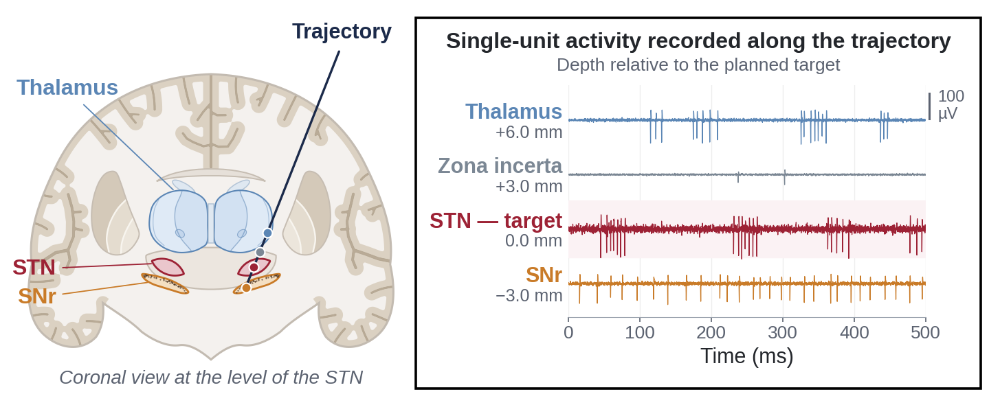
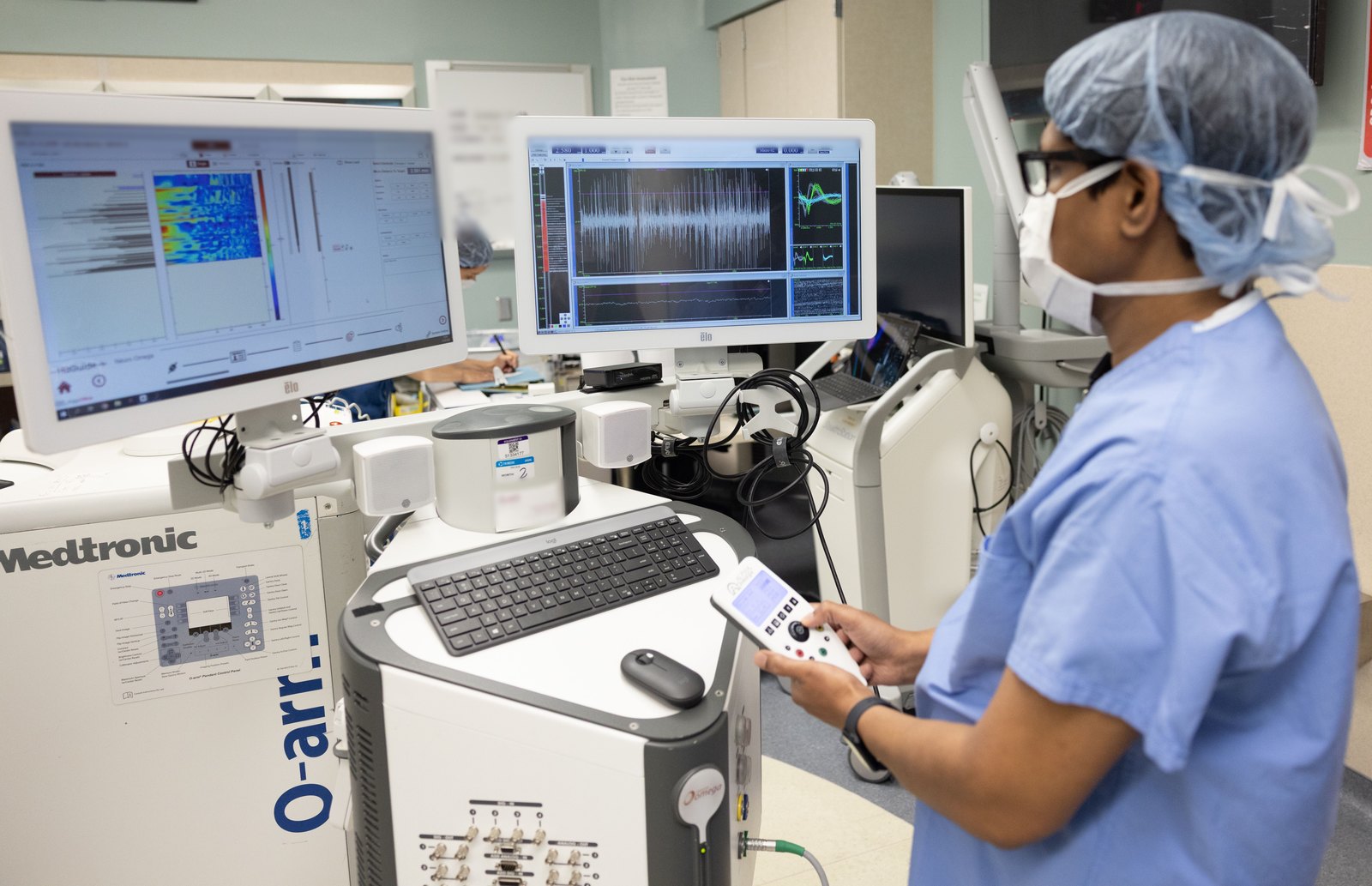
Microelectrode recording is performed for target mapping during deep brain stimulation (DBS) surgery and allows the activity of individual neurons and neuronal populations to be recorded from deep brain structures. We use these recordings to investigate patterns of neuronal activity and examine how they relate to brain function and neurological disorders.
Some DBS devices have sensing capability, meaning they can record local field potentials longitudinally from the patient’s brain while they go about their daily lives. These chronic recordings provide opportunities to study how neural activity changes over time and across disease, behavioral, and treatment states. We use these recordings to identify clinically meaningful neural biomarkers that may help characterize symptoms, track treatment response, and inform the development of more personalized DBS therapies.
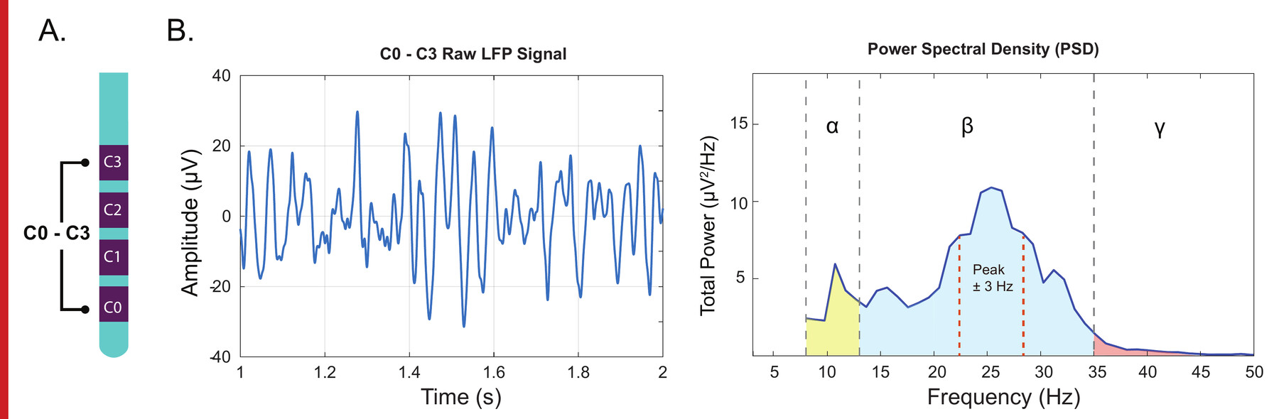
Another theme is using computational and biophysical modeling to better understand neural recordings and the effects of brain stimulation. We develop models that integrate neurophysiology with patient-specific anatomy to investigate where recorded neural signals originate and how electrical stimulation interacts with neural tissue. These approaches help us interpret signals recorded from implanted electrodes, link measured brain activity to its underlying neural sources, and provide mechanistic insights that complement our experimental studies.
Human recordings can identify candidate neural circuits and biomarkers, but many causal mechanisms cannot be tested directly in patients. In collaboration with basic scientists, we perform experiments in animal models using approaches including optogenetics to manipulate specific neural circuits and test their roles in behavior and disease. These mechanistic studies complement our human neurophysiology and modeling work and help determine how specific circuits may be targeted through neuromodulation.
Together, these research approaches create a framework for precision neuromodulation: direct human brain recordings help identify disease-relevant neural activity and circuits; chronic recordings reveal how these signals evolve over time; computational and biophysical models help interpret neural signals and understand how stimulation engages the brain; and mechanistic experiments test causal circuit hypotheses. The long-term goal is to translate this knowledge into neuromodulation therapies tailored to the neural circuitry and physiology of individual patients.